


Osteochondrosis is an outdated name for a degenerative spine disease. The old term is widely used in our country, but does not reflect the essence of the disease, which is based on age-related degeneration - destruction of tissue structure. In this article we will consider the first signs of osteochondrosis, its development patterns and treatment options.
What is Osteochondrosis?
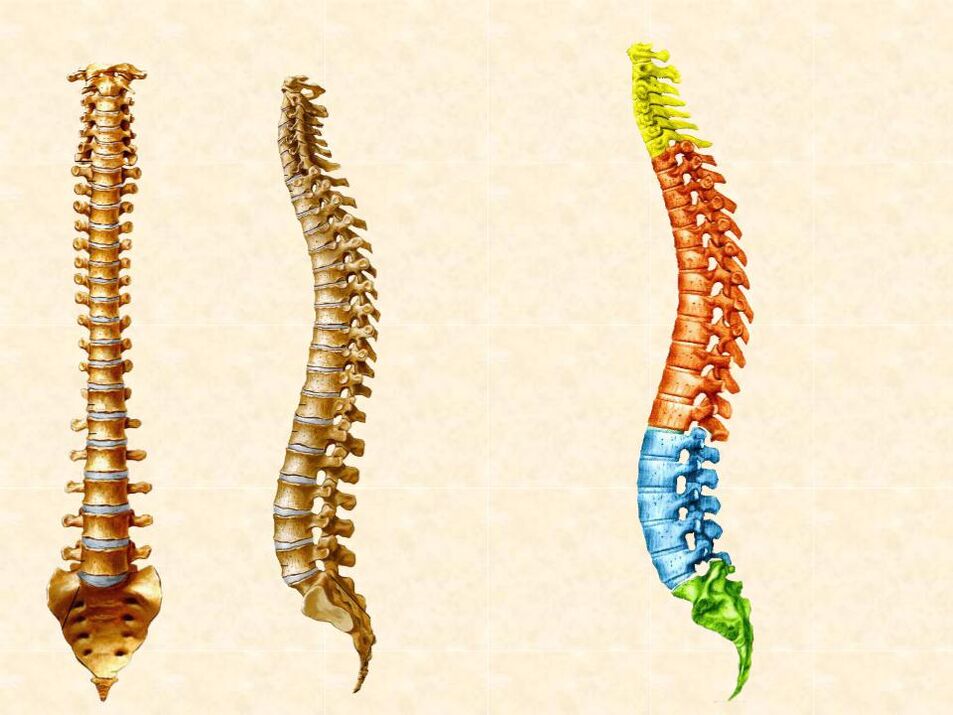
To understand the processes occurring in osteochondrosis, you need to understand the anatomy of the spine. It includes the following structures:
- Vertebrae consisting of bodies, arches, processes. Between the arches of adjacent vertebrae there are joints called facets
- Intervertebral discs located between the bodies of adjacent vertebrae
- Spinal ligaments
- The posterior and anterior longitudinal axes run along the bodies of all vertebrae at the front and back
- Ligamentum flavum – connects the arches of adjacent vertebrae
- Supraspinous ligaments and interspinous ligaments – connect the spinous processes
- The spinal cord, which is located in the spinal canal, along with the nerve roots radiating from it. They are extensions of nerve cells. Through these processes, the brain receives information about the state of tissues and, in response, sends signals that regulate their function: muscle contractions, changes in blood vessel diameter, and much more.
Degeneration begins with the intervertebral discs and as the change progresses, all of the structures mentioned above become involved in the process. This is partly because the discs have no blood vessels. Nutrients and oxygen enter them by diffusion from the vertebrae and other surrounding structures.
Intervertebral discs make up a third of the length of the spine and serve as shock absorbers. They protect the vertebrae from overloading when lifting heavy loads, standing or sitting for long periods of time, and when bending and twisting. Each hard drive consists of:
- The nucleus pulposus, located inside, in the middle, contains a lot of hyaluronic acid, collagen type II, which stores water. This gives the normal core a jelly-like consistency for effective steaming. As degeneration progresses, the composition of the inner part of the intervertebral disc changes, its water content decreases, the core "dries out" and the height of the intervertebral disc decreases
- The fibrous ring, which is located outside the cell nucleus and consists of 15-25 layers of collagen fibers. Collagen in the annulus fibrosus is type I. It is denser than in the nucleus and is needed to hold the interior of the disc and protect it from damage. The fibers of the ring are intertwined along the periphery with the posterior longitudinal ligament of the spine. This ensures the immobility of the spinal structures in a healthy person - doctors call this condition spinal stability. In people with degenerative disease, the annulus fibrosus tears, which can lead to instability: adjacent vertebrae can move forward or backward relative to each other. This is dangerous because the nerve root becomes trapped between them
It is also important to mention the end plates. These are thin cartilages that are located between the vertebral bodies and intervertebral discs. They contain blood vessels that supply the intervertebral disc. In degenerative diseases, calcium builds up in the end plates, resulting in impaired blood supply.
Stages of development of osteochondrosis
The development of spinal osteochondrosis occurs gradually:
- Initial degeneration. The intervertebral disc does not receive sufficient nutrition, wears out, its height decreases and cracks occur. The nucleus pulposus protrudes due to microdamage to the annulus fibrosus, irritating the posterior longitudinal ligament and leading to pain and reflex spasms of the back muscles
- Bulging of the intervertebral disc. The fibers of the annulus fibrosus are destroyed, the nucleus pulposus becomes more prominent and a hernia develops. It can compress the roots of the spinal nerve, leading to the development of paresis or paralysis of the limb muscles and a reduction in skin sensitivity. One of the complications of a hernia is its sequestration - the separation of the disc protrusion from its main part.
- Progressive degeneration of the prominence and other structures of the spine. The intervertebral disc becomes even more compact and the body tries to compensate for the excessive mobility of the spine by forming pathological bone growths of the vertebral bodies - osteophytes. Like the hernia itself, they can affect nerves and ligaments, impairing their function and causing pain. Unlike a hernia, bone spurs do not dissolve.
Complications of osteochondrosis, in addition to compression of the herniated spinal nerve roots:
- Spondyloarthrosis. Reduced disc height results in greater stress on the facet joints. They can develop inflammation and malnutrition, causing them to become "dry" and cause pain.
- Spondylolisthesis 一 Displacement of the vertebral bodies relative to each other due to damage to the ligaments
- Degenerative processes in the area of the ligamentum flavum lead to its thickening. This is dangerous because the yellow ligament is adjacent to the spinal canal and can narrow it and compress the spinal cord
- At the height of the 1st -2ndlumbar vertebra, it extends downwards from the spinal cord"ponytail" – a bundle of nerve roots responsible for the innervation of the lower extremities and pelvic organs: bladder, rectum, external genitals. Cauda equina syndrome is one of the most dangerous complications of osteochondrosis and is manifested by severe pain, muscle weakness in the legs, numbness in the perineum and urinary and fecal incontinence.
Causes of osteochondrosis of the back
There is still no consensus about what degree of degenerative changes in the spine should be considered normal. Sooner or later, everyone's spine begins to age.
For most people, these changes are minor and do not cause any symptoms: sometimes they are discovered accidentally during a magnetic resonance imaging (MRI) scan of the spine. The progression of degeneration leads to significant changes in the structure of the spine. Intervertebral discs can become so destroyed that they no longer perform their shock-absorbing function, bulging and putting pressure on the spinal nerves and even the spinal cord itself.
It is impossible to predict exactly how severe the degenerative changes will be in a particular person and whether they will lead to complications. There is a genetic predisposition to osteochondrosis, but the specific genetic mutations responsible for the disease progression have not been identified. Therefore, there is no precise genetic test that would show personal risk. There are certain factors that increase the risk of osteochondrosis. It is they who are targeted by measures to prevent osteochondrosis.
Risk factors for osteochondrosis include:
- Excessive stress on the spine: Professional sports, heavy lifting, regular heavy physical work
- Prolonged stay in a static, incorrect position: sitting, slouching, cross-legged, on a chair without lumbar support, working in a vertical position with an inclination
- Sedentary lifestyleThis leads to weakness in the core muscles, which cannot effectively support the spine
- Overweight Excess weight puts additional strain on the back and joints
- Smoke - Nicotine and other components of cigarettes interfere with the diffusion of nutrients from blood vessels into tissues, including intervertebral discs
- Alcohol consumption - Regular consumption leads to poor absorption of calcium from food. A lack of calcium causes the vertebrae to lose density
- Back injuries with damage to the structure of the vertebrae or intervertebral discs, as a result of which the recovery process is much slower than the degeneration process
Osteochondrosis of the spine in adults: symptoms
In the early stages of a degenerative disease, a person usually does not experience any symptoms. They appear suddenly or gradually as the disease progresses. The main symptoms are back pain and reflex spasms of the back muscles. The location of the symptoms depends on which part of the spine the problem occurs:
- Degeneration of the cervical spine causes muscle stiffness, neck pain that radiates to the shoulder and arm or back of the head, and worsens with head movement
- Changes to the thoracic spine occur extremely rarely because it is the most static. If a hernia does occur, pain occurs between the shoulder blades
- Hernias in the lumbar region occur more often than others and are manifested by pain in the lower back or sacrum that radiates to the gluteal region and leg. Stiffness in the lower back is also noted. The pain increases when sitting, standing for long periods of time and bending over
If pain radiates from the back into the extremities, it is referred to as radiculopathy - damage to the nerve root. This is compression caused by a herniated disc of the spinal nerve. In addition to pain, radiculopathy is accompanied by other symptoms that are localized to a specific area supplied by the damaged nerve. These manifestations may include:
- Weakness of the muscles of the limbs up to paralysis
- Disturbances in the sensitivity of the skin of the extremities
- Bladder and rectal dysfunction with lumbar radiculopathy
The signs of spinal osteochondrosis generally do not differ in women and men, but in women, symptomatic degeneration develops more quickly after menopause when bone density decreases. In men, degenerative processes are more often caused by physical work and develop at an earlier age, but gradually.
Not all back pain is caused by spinal osteochondrosis. Our specialists can perform a full examination and decide whether you need an MRI.
Osteochondrosis of the spine at a young age
It is generally accepted that osteochondrosis is a disease of the elderly. While degenerative spine disease is common in patients over the age of 60, it is becoming increasingly common in people in their 30s and even 20s. The cause is usually genetics, obesity, lack of exercise or back injuries. Both one-off serious injuries, for example due to a fall, and regular minor injuries, for example in professional sports, are important. The condition most commonly occurs in the lumbar region because it is the most mobile. Intervertebral hernias, including Schmorl nodes, can form here. The main mechanism of their occurrence is damage to the endplates, which cannot withstand intradiscal pressure. In this way, protrusions arise in the body of the vertebra above or below, so-called Schmorl hernias. They do not cause compression of the nerve roots and are generally harmless. In rare cases, they can grow and cause back pain, but more often they are discovered accidentally during an MRI. Backwardly protruding intervertebral hernias are usually associated with pain and may require treatment.
Osteochondrosis of the spine: treatment
Up to 90% of degenerative diseases can be treated with conservative methods.
Surgery is only indicated if there is a risk of serious complications, such as progressive loss of bladder control or weakness of the lower extremities. Surgical treatment allows you to save a person from paralysis, but does not relieve pain and further progression of the disease. Therefore, a special rehabilitation program is prescribed after the operation.
In many cases, uncomplicated hernias resolve on their own. The resorption process may be accompanied by the formation of excess connective tissue and calcifications in the spine, increasing the likelihood of recurrence of the disease in the future. Existing physiotherapeutic techniques and special exercises help:
- accelerate the resorption of the hernia
- Improve disk performance
- Normalization of the biomechanics of movements and load distribution
- Avoid the need for surgery in the future
Medications from the groups of non-steroidal anti-inflammatory drugs, glucocorticoids and muscle relaxants are also used to combat pain, but the use of medication is limited to the acute phase of the disease and does not lead to a permanent improvement in the condition of the spine. You can reduce the intensity of degeneration by:
- MLS laser therapy – the laser radiation used has an anti-inflammatory effect, expands the lymphatic vessels and improves lymphatic drainage
- Acupuncture – this method relieves pain, swelling and inflammation due to the body's reflex reaction to stimulation of biologically active points on the body with special needles
- The magnetotherapy method stimulates blood circulation, normalizes the diffusion of nutrients and removes toxins from the thickness of the intervertebral discs, thereby accelerating the recovery processes
- Therapeutic physical education - special sets of exercises help to strengthen the core muscles, learn the correct distribution of load on the back, maintain correct posture and relieve muscle spasms. To monitor performance, it is better to work with an instructor and then continue the exercises yourself according to the recommendations
Depending on the severity of the disease and the characteristics of the patient, different combinations of the above methods can be used.
Both conservative treatment of spinal hernias and rehabilitation after operations can be carried out on an outpatient basis in the clinic. It has all the necessary equipment and a team of professionals specialized in non-surgical hernia treatment. It is not recommended to go to hospitals that use methods that are not scientifically based and not recognized by the world medical community - this can be dangerous to health. In a modern clinic you can get advice at low cost and determine the next steps together with your doctor.